
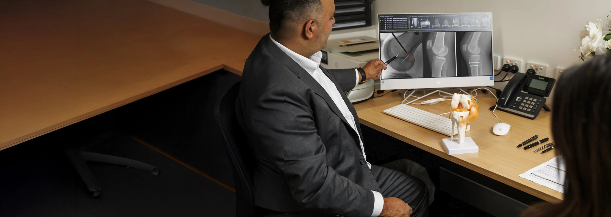
Patella Realignment Surgery
Patella realignment surgery treats recurrent instability or maltracking of the kneecap (patella) when the cause lies partly in the shape or alignment of the bones around the knee. It is usually combined with MPFL reconstruction.
Dr Varun Arora performs patella realignment surgery for patients across Melbourne and regional Victoria with ongoing kneecap instability that has not responded to non-surgical treatment.
What is patella realignment surgery?
Kneecap instability usually reflects a combination of a torn or stretched ligament (the MPFL) and anatomical factors, such as a shallow groove, a high-riding kneecap or a tendon that pulls the kneecap outwards. Realignment surgery corrects these bony factors so the kneecap sits and tracks securely in its groove. Depending on the cause, it may involve soft-tissue procedures, bony realignment, or a combination of both.
1. Patella Instability → causes, first dislocations and your options
Kneecap Instability Guide
2. MPFL Reconstruction → rebuilding the ligament that holds the kneecap
3. Patella Realignment (this page): correcting the bony factors behind instability
Is patella realignment right for me?
Realignment is usually recommended when your kneecap keeps dislocating and your scans show one or more anatomical factors that MPFL reconstruction alone would not correct:
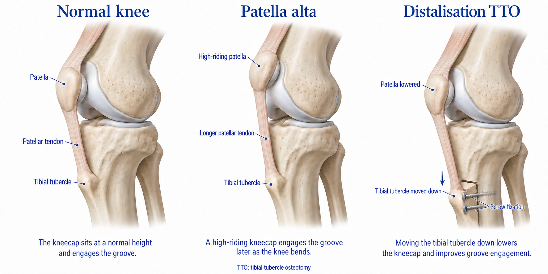
A high-riding kneecap (patella alta)
A large TT–TG distance, where the patellar tendon pulls the kneecap outwards
Severe trochlear dysplasia, where the groove is flat or bulging
Significant twisting of the thigh bone or shin bone
Instability that has returned after previous kneecap surgery
Surgery works best when it addresses the specific factors present in your knee. Treating only one of several causes increases the risk of instability returning. These factors are explained on Patella Instability.
Matching the procedure to the cause
Most people need only one or two of these procedures. MPFL reconstruction is the most common, and the others are added when your anatomy calls for them.
| Procedure | Underlying cause it treats | What it involves |
|---|---|---|
| MPFL reconstruction | A torn or stretched MPFL. Present in almost everyone with recurrent dislocation. | Rebuilding the ligament with a tendon graft. Often enough on its own when the rest of the anatomy is close to normal. See MPFL Reconstruction. |
| Tibial tubercle osteotomy: medialisation | A large TT–TG distance, pulling the kneecap outwards | The bony bump where the tendon attaches is cut, moved inwards and fixed with screws |
| Tibial tubercle osteotomy: distalisation | A high-riding kneecap (patella alta) that doesn’t sit securely in the groove | The same bony bump is moved downwards to lower the kneecap. It can be combined with medialisation. |
| Trochleoplasty | Severe trochlear dysplasia, where the groove is flat or even bulging | Reshaping and deepening the groove at the end of the thigh bone. Performed in carefully selected cases, usually with MPFL reconstruction. |
| Lateral lengthening | Tight tissues on the outer side of the knee that tilt the kneecap and pull it outwards | Lengthening the tight outer tissue. Used as an addition to other procedures, not as a treatment for instability on its own. |
| Derotation osteotomy | Significant twisting of the thigh bone or shin bone, which changes the direction the kneecap is pulled | Cutting the bone, rotating it into a better position and fixing it with a plate or rod. Reserved for marked rotational deformity. |
Tibial tubercle osteotomy explained
The tibial tubercle is the bony bump just below the knee where the patellar tendon attaches. Moving it changes the direction and height of the pull on the kneecap. Through an incision at the front of the shin, a strip of bone carrying the tendon attachment is cut, shifted inwards, downwards or both by a planned amount, and fixed with screws. It heals like a fracture, which is why weight-bearing is protected for the first few weeks.
Lateral release on its own
In the past, a lateral release (cutting the tissues on the outer side of the kneecap) was commonly performed by itself for kneecap instability. It is no longer recommended as a stand-alone treatment for instability, because it doesn't address the cause and can occasionally make the kneecap unstable towards the inner side.
Children and teenagers
Bony procedures such as tibial tubercle osteotomy and derotation osteotomy are generally delayed until growth has finished, because they can affect the growth plates. Teenagers who are still growing usually have MPFL reconstruction alone, using growth-plate-friendly techniques. See MPFL Reconstruction.
What is involved in patella realignment surgery?
Before surgery
Examination and imaging, usually x-rays and an MRI and sometimes a CT scan, measure the factors contributing to instability: the shape of the groove, the height of the kneecap, the TT–TG distance and leg rotation. These measurements guide the combination of procedures planned for you.
Arthroscopy
The knee is usually inspected with a camera first. Any loose fragments are removed, and the cartilage behind the kneecap is checked.
Bony realignment
Where needed, the tibial tubercle is repositioned and fixed with screws to improve the alignment and height of the kneecap. Less commonly, the groove is deepened or a rotational deformity is corrected.
Soft-tissue stabilisation and testing
The MPFL is usually reconstructed with a tendon graft and, where needed, tight outer tissues are lengthened. The kneecap's tracking is then tested through a full range of motion before the incisions are closed.
Your recovery
Recovery depends on the combination of procedures performed. It generally involves a period of bracing and protected weight-bearing, followed by a structured physiotherapy program.
Recovery at a glance
Soft-tissue surgery alone recovers fastest. Bony procedures need extra time for the bone to heal.
-
Day 0–1
Up with crutches and a hinged brace. Most go home the same day or the next day.
-
0-6 weeks
After tibial tubercle osteotomy: protected weight-bearing on crutches while the bone heals
-
at around 6 weeks
X-ray check, then increasing weight and weaning off crutches. Protected weight-bearing after a derotation osteotomy or trochleoplasty usually continues to about this point.
-
6-8 weeks
Driving and desk-based work, depending on the procedure and which knee -
3 months
The bone is usually healed. Cycling, swimming and progressive strengthening.
-
around 4 months
Straight-line running, if strength and control goals are met
-
6-9 months
Return to pivoting and contact sport once criteria are met
If you have MPFL reconstruction alone, see the faster timeline on MPFL Reconstruction. See also Post-operative Recovery & Rehab Protocols.
Returning to sport
Return to sport after realignment is guided by the same milestones as after MPFL reconstruction: full, comfortable movement, strength close to the other leg, good hopping and landing control, and confidence in the kneecap. Because bone has to heal first, these milestones are usually reached later, at around six to nine months.
General risks overview
IMPORTANT
As with any surgical procedure, patella realignment surgery carries risks. Potential risks and complications include:
Infection
Blood clots (deep vein thrombosis or pulmonary embolism)
Recurrent instability
Stiffness
Ongoing pain around the kneecap
Over-correction, which can increase pressure behind the kneecap or cause instability to the inner side
Risks specific to bony realignment, such as delayed bone healing, fracture of the shin bone, or irritation from screws that may need later removal
Nerve or blood vessel injury
Risks associated with anaesthesia
This is a general overview and not a complete list. Dr Arora will discuss the risks and benefits relevant to you in detail before surgery.
Patella Realignment Frequently Asked Questions
Is it right for me?
-
MPFL reconstruction rebuilds the ligament that holds the kneecap. Realignment is a broader term that also includes bony procedures, used when the shape or alignment of the bones is contributing to your instability. The two are often combined. See MPFL Reconstruction.
-
Dr Arora will assess the underlying cause of your instability through examination and imaging, and recommend the most suitable combination. See the table above.
-
A lateral release on its own is not recommended for kneecap instability, as it doesn't address the cause and can make the kneecap unstable in the other direction.
-
Bony realignment is generally delayed until growth is complete. Teenagers who are still growing can usually have MPFL reconstruction with growth-plate-friendly techniques.
-
Yes. Revision surgery is planned after a careful review of what is still causing the instability, with up-to-date imaging. Often a bony factor that wasn't addressed the first time is corrected.
-
Yes. When both hips need replacing, the anterior approach can be used on each side, either together or a few months apart.
The operation
-
The bony bump below the knee where the patellar tendon attaches is cut, moved to a better position and fixed with screws. This changes the direction or height of the pull on the kneecap.
-
An operation to deepen a flat or bulging groove at the end of the thigh bone. It is reserved for severe groove dysplasia in carefully selected patients who have finished growing.
-
Often, yes. MPFL reconstruction is commonly combined with a tibial tubercle osteotomy or, less often, other procedures in the same operation.
-
Usually not. If they are prominent and uncomfortable, especially when kneeling, they can be removed once the bone has fully healed.
Recovery
-
It depends on the procedures performed. After bony realignment, return to sport is usually at six to nine months. After MPFL reconstruction alone, it is usually four to six months.
-
Usually around six weeks after a bony procedure, and two to four weeks after soft-tissue surgery alone.
-
A hinged brace is usually worn for the first four to six weeks. It protects the repair while movement returns.
-
Usually six to eight weeks after bony realignment, depending on which knee was treated and the type of work you do. Physical work may take three months or more.
The long term
-
The risk is low when surgery addresses the factors causing your instability, but no surgery can guarantee it will never happen again.
-
Most people can kneel, although it may be uncomfortable for many months, particularly after a tibial tubercle osteotomy.
-
Occasionally, if screws or a plate remain. You don't need a letter. Just let security staff know.
Trusted resources
These independent sources give more information about kneecap instability and realignment surgery.
PATIENT INFORMATION
PROCEDURE INFORMATION
External links are provided for general information. Dr Arora is not responsible for the content of external websites.
General information only. Any surgical or invasive procedure carries risks. Before proceeding, you should seek a second opinion from an appropriately qualified health practitioner.

Knee Surgery in Melbourne
To discuss ongoing kneecap instability and your treatment options, call (03) 9088 8420 or contact Dr Varun Arora to schedule a consultation. A GP referral is required.