
Patella Instability
Patella instability is when the kneecap (patella) slips partly or completely out of its groove at the end of the thigh bone. It is most common in teenagers and young adults, often during sport or a twisting movement.
Dr Varun Arora assesses and treats patients with kneecap instability across Melbourne and regional Victoria, from a first dislocation in a teenager to recurrent instability that limits sport and daily life.
What is patella instability?
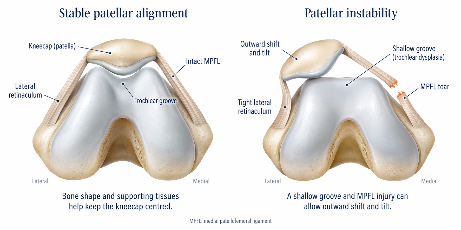
A complete slip of the kneecap out of its groove is a dislocation, and a partial slip is a subluxation. The kneecap almost always moves towards the outer side of the knee, and it often pops back into place by itself. The kneecap is held in place by the shape of its groove and by soft tissues, especially the medial patellofemoral ligament (MPFL) on the inner side of the knee. The MPFL is almost always torn or stretched when the kneecap dislocates.
1. Patella Instability (this page): causes, first dislocations and your options
Kneecap instability guide
2. MPFL Reconstruction → rebuilding the ligament that holds the kneecap
3. Patella Realignment → correcting the bony factors behind instability
Symptoms
Common symptoms of patella instability include:
The kneecap visibly shifting to the outer side of the knee, often with a twisting movement
Sudden pain and swelling after the kneecap dislocates
A feeling that the kneecap is about to slip, especially when twisting or changing direction
The knee giving way
Pain around or behind the kneecap
Avoiding sport or certain movements because you don't trust the knee

Causes and risk factors
Most people with kneecap instability have one or more anatomical features that make the kneecap easier to dislocate. These are measured on x-rays, MRI and sometimes CT, and they guide treatment:
Trochlear dysplasia. A shallow, flat or bulging groove at the end of the thigh bone, so the kneecap has less bony support
Patella alta. A high-riding kneecap that engages the groove later as the knee bends
A raised TT–TG distance. The patellar tendon attaches further to the outer side of the shin bone, pulling the kneecap outwards
Leg alignment and rotation. Knock knees or twisting of the thigh or shin bones
Ligament laxity. Generally loose or "double-jointed" ligaments
Age and previous dislocation. Being young, especially while still growing, and having dislocated before
Straight after a dislocation
-
If the kneecap is still out of place, go to an emergency department.
Don't try to force it back yourself.
-
Once it is back in place, settle the knee.
Rest, ice, a support or brace, and crutches if you are limping.
-
See your GP within a few days.
They can examine the knee and arrange an x-ray and MRI.
-
Start physiotherapy
To settle swelling, restore movement and begin strengthening.
-
Arrange a surgical review
If the MRI shows a loose fragment, the knee stays swollen or locks, or this is not your first dislocation.
How is patella instability diagnosed?
Diagnosis is based on your history and an examination of the kneecap's movement and stability. Imaging then looks at two things: damage from the dislocation, and the anatomical factors that made it happen.
X-rays look for fractures and show the shape of the groove and the height of the kneecap.
MRI is usually recommended after a dislocation. It checks for cartilage damage or loose fragments and measures the risk factors above.
CT is occasionally used to assess bone alignment and rotation before surgery.
Will it happen again?
After a first dislocation, around one in three people have another one. The risk is much higher in younger patients, those who are still growing, and those with trochlear dysplasia, patella alta or a raised TT–TG distance. With several of these factors, a further dislocation becomes more likely than not. Assessing these factors helps decide who may benefit from earlier surgery.
Treatment options for patella instability
Treatment depends on whether this is your first dislocation or a recurrent problem, whether there is damage inside the joint, and your individual anatomy.
After a first dislocation
Most first dislocations are treated without surgery:
Settling the knee: a short period in a brace or support, with crutches if needed
Physiotherapy: restoring movement, then strengthening the quadriceps, hip and core muscles that control the kneecap
A gradual return to sport, sometimes with taping or a brace for confidence
Early surgery is recommended if the MRI shows a loose fragment of bone or cartilage that needs to be fixed or removed. It may also be considered for patients with several high-risk features.
Surgery for recurrent instability
When the kneecap keeps dislocating, or instability is limiting your life despite physiotherapy, surgery is usually recommended. The operation is matched to the factors causing your instability.
| Comparison | MPFL reconstruction alone | MPFL reconstruction with bony realignment |
|---|---|---|
| What it does | Rebuilds the torn ligament with a tendon graft, acting as a “check rein” for the kneecap | Rebuilds the ligament and also corrects the bony factors, most often by moving the tendon attachment on the shin bone (tibial tubercle osteotomy) |
| Usually suits | Recurrent dislocation with near-normal anatomy. This is the most common operation. | A high-riding kneecap, a raised TT–TG distance, severe groove dysplasia or marked rotation |
| Return to sport | Usually 4–6 months | Usually 6–9 months |
| Children still growing | Possible, using growth-plate-friendly techniques | Bony procedures usually wait until growth is complete |
| More information | MPFL Reconstruction | Patella Realignment |
Children and teenagers
Kneecap instability often starts in the teenage years, and young age is one of the strongest risk factors for further dislocations. A first dislocation in a child is usually treated with physiotherapy, unless there is a loose fragment. If dislocations keep happening, MPFL reconstruction can be performed while the child is still growing, using techniques that protect the growth plate. Bony realignment is usually delayed until growth has finished.
When should I see a surgeon?
Consider asking your GP for a referral if:
Your kneecap has dislocated for the first time, especially if the knee remains swollen or locks
Your kneecap has dislocated more than once
You feel the kneecap slipping, or don't trust the knee, despite physiotherapy
Your child or teenager has had a kneecap dislocation
Seeing a surgeon doesn't commit you to an operation. Many patients come for a clear diagnosis and a plan.
Patella Instability Frequently Asked Questions
Understanding patella instability
-
In a dislocation, the kneecap comes completely out of its groove. In a subluxation, it slips partly out and returns. Both are forms of patella instability.
-
Most people with kneecap instability have one or more anatomical factors, such as a shallow groove, a high-riding kneecap or loose ligaments, that make the kneecap easier to dislocate with a twisting movement.
-
It is a measurement on CT or MRI of how far to the outer side the patellar tendon attaches on the shin bone. A large distance pulls the kneecap outwards and can contribute to instability.
-
Not necessarily. Pain around the kneecap without it slipping (patellofemoral pain) is common and is treated differently, usually with physiotherapy. An assessment helps tell the two apart.
-
Yes. The anatomical factors behind instability are often present in both knees, although usually only one causes problems.
After a first dislocation
-
Yes. Even when the kneecap relocates on its own, the dislocation can chip off fragments of cartilage or bone. An assessment and usually an MRI are recommended.
-
Around one in three people have another dislocation after the first. The risk is higher in young patients and those with certain anatomical features, which Dr Arora can assess for you.
-
Usually not. Most first dislocations are treated with physiotherapy. Surgery is recommended early if there is a loose fragment in the joint, and may be considered sooner for patients with several high-risk features.
-
Many people return to sport over 6 to 12 weeks, once swelling has settled and strength and control have returned.
-
A brace or taping can help with confidence in the early weeks and when returning to sport, but strengthening is the most important part of treatment.
-
Strengthening the quadriceps, hip and core muscles, along with balance and landing control guided by a physiotherapist, is the foundation of treatment.
Choosing a treatment
-
Usually when the kneecap has dislocated more than once, or still feels unstable despite physiotherapy. It is also recommended earlier if there is a loose fragment in the joint.
-
Most people need MPFL reconstruction alone. Bony realignment is added when scans show a high-riding kneecap, a raised TT–TG distance or other significant anatomical factors. See the table above.
-
Yes. Children and teenagers who are still growing can have MPFL reconstruction using techniques that protect the growth plate. Bony procedures usually wait until growth is complete.
-
Repeated dislocations can damage the cartilage behind the kneecap and increase the risk of arthritis later. Stabilising a knee that keeps dislocating helps protect it.
Trusted resources
These independent sources give more information about kneecap instability.
Patient information
INJURY PREVENTION
FURTHER READING: THE EVIDENCE
External links are provided for general information. Dr Arora is not responsible for the content of external websites. General information only. Any surgical or invasive procedure carries risks. Before proceeding, you should seek a second opinion from an appropriately qualified health practitioner.

Knee Surgery in Melbourne
If your kneecap has dislocated or feels unstable, call (03) 9088 8420 or contact Dr Varun Arora to schedule a consultation. A GP referral is required.